#Auditory cortex damage full
In addition, the results also point to the capacity of each telencephalic hemisphere to process the full range of auditory lateralization from left to right. The results indicate the specificity of the dichotic signal/noise tests for the identification of unilateral lesions in thalamocortical auditory structures. auditory cortex on each side of the brain is sensitive to.

All of them showed unimpaired performance in all test alternatives. potential for broad patterns of damage on the cellular level in addition to well-defined. Another 24 patients were studied who had lesions mostly close to but sparing the before-mentioned auditory structures.

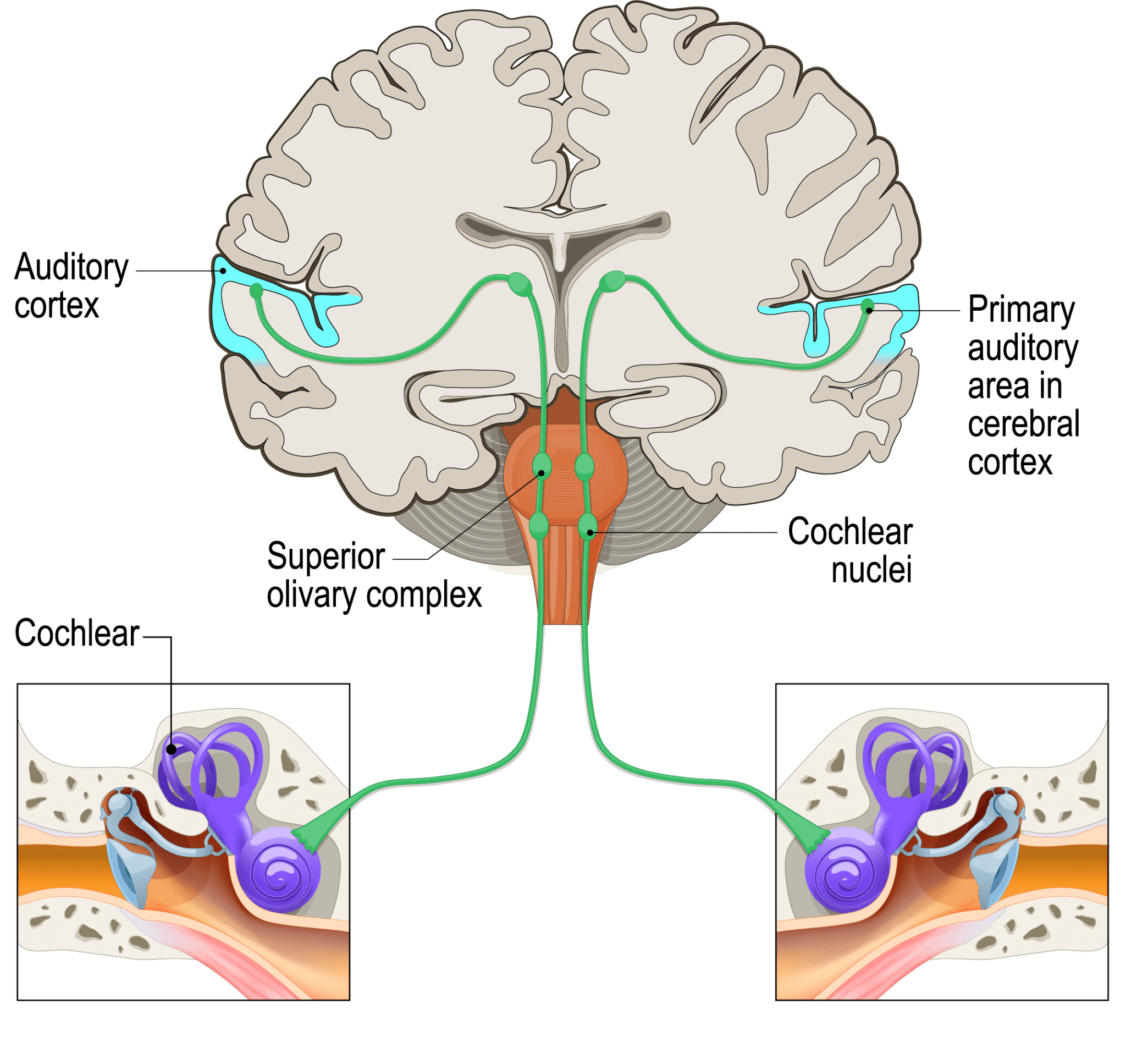
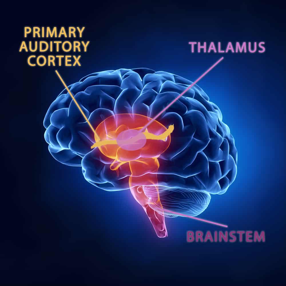
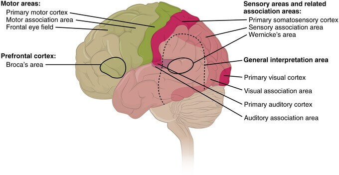
All patients were able to master the interaural tests, which indicates the preserved ability to lateralize sound sources to the left and to the right with either one of the auditory cortices left intact. With inverted signal and noise stimulation, however, the thresholds were in the range of age-matched controls. The patients showed impaired discrimination of signal frequency, intensity and duration in the dichotic signal/noise tests, when the signals were presented to the ear contralateral and the noise ipsilateral to the lesion. Updated on FebruReviewed by Saul Mcleod, PhD The cerebral cortex is the brain’s outermost layer that lies on top of the cerebrum and is associated with our highest mental capabilities. Sensorineural, which involves the inner ear. Cortical deafness is an auditory disorder. There are three types of hearing loss: Conductive, which involves the outer or middle ear. Cortical deafness is a rare form of sensorineural hearing loss caused by damage to the primary auditory cortex. More than half the people in the United States older than age 75 have some age-related hearing loss. Monaural tests of either ear revealed no deficits in auditory performance. Bilateral damage to the auditory cortex is not associated with permanent deficits in the ability to detect sound. Overview Hearing loss that comes on little by little as you age, also known as presbycusis, is common. Location and extent of the lesions were assessed by magnetic resonance imaging. For example, when sensory cells in the inner ear are damaged from loud noise, the resulting hearing loss changes some of the signals in the brain to cause tinnitus. Twenty-one patients from neurology were studied who suffered from unilateral lesions in the auditory cortex, the auditory thalamus, or the acoustic radiation. Something as simple as a piece of earwax blocking the ear canal can cause tinnitus, but it can also arise from a number of health conditions. The different test alternatives generated distinct auditory percepts, which is in accordance with the assumption of specific signal processing at the level of the auditory brainstem and at thalamocortical auditory areas. The auditory cortex in the temporal lobe is key for hearing and.

Thresholds for the discrimination of signal frequency, intensity and duration were acquired under three different conditions of headphone stimulation (‘monaural’, ‘interaural’, and ‘dichotic signal/noise tests’) using the three-alternative forced-choice procedure. Damage to this region of the brain can have global consequences for virtually every. The extent of perceptual impairment following unilateral lesions in the auditory cortex, its thalamic or callosal afferents was studied with psychoacoustic tests.


 0 kommentar(er)
